‘Oderus’ The Super Dog
Oderus, a 19 week old male German Shepherd puppy, presented to the Animal Emergency Clinic of the Fraser Valley for progressive ataxia(falling over) and muscle stiffness. Symptoms had been getting worse over the previous two days and a decrease in appetite for five days. On exam he was exhibiting sings of anxiety, sound/light sensitivity and ataxia with muscle and limb rigidity, looking like a saw horse at times when stimulated. He also had difficulty swallowing and looked as though he was foaming at the mouth. Within the first six hours of admittance Oderus could not stand or walk on his own and his respiratory defenses became compromised.
The main goal of treatment for Tetanus is, one; find the source of infection and remove it, two; bind the remaining toxin in the system by administering antitoxin and three; provide supportive care to the patient while recovering.
Oderus received around the clock care consisting of changing his body position to prevent bed sores and lung atelectasis (collapse), nutritional supplementation via intravenous and feeding tubes because he could not swallow on his own, oxygen and humidification to increase oxygen saturation, antibiotics to combat any secondary infections, tetanus antitoxin to bind the remaining toxin, gastric protectants, pain medications and sedation/muscle relaxation to alleviate stress and suffering.
We found the source of infection for Oderus to be one of his deciduous (baby) teeth and removed the infected tooth while at the same time, placing a feeding tube called a ‘PEG Tube’ directly into his stomach for nutritional support. Oderus was in our care for two weeks. When we sent him home he was able to walk with the help of his parents, was eating food on his own and received the remainder of his caloric requirements via his feeding tube. With his parents diligent care we were able to remove his feeding tube within a week of being sent home and soon after, Oderus was swimming and building his strength back. He is now a big happy, healthy and boisterous super dog who still loves to come back to the clinic for visits.
Without treatment Oderus would not have survived. His parents’ commitment to giving him the time required to recover and heal from the tetanus toxoid injury along with continuous nursing care was the key to his recovery.
Tetanus
A case base approach
Tetanus can occur following an infection of deep wounds that have been contaminated by the bacteria clostridium tetani. When thinking of tetanus you might think of the rusty nail being stepped on when in fact the bacteria is commonly found in soil and in the intestinal tract of mammals as part of normal flora which is why some animals can contract tetanus after having been payed or neutered. Clostridium Tetani only becomes a problem when there is a break in the natural barriers, ex. skin, and bacteria enters the deep body tissues where it begins to produce a toxin which attacks the nervous system by preventing transmission of nerve impulses. As nerve transmissions are blocked, the whole body may then become very reactive to touch, light and sound. In addition, the respiratory system can also become affected directly, from muscle paralysis, and or indirectly through aspiration of oral secretions or food and water.
Tetanus, though rare in small animal medicine, is more common in dogs than in cats as cats appear to be more resistant to the infection than dogs. The clinical signs may be seen as early as four days and as far as eight weeks . A quicker onset of signs may indicate a poorer prognosis.
Affected animals at home may show acute (sudden onset) generalized stiffness, lethargy, light/sound sensitivity and difficulty eating due to muscle rigidity of the jaw. A classic tetanus sign is the joker grin that results from contraction of the muscles in the jaw, face and head. If left untreated the disease can progress to more severe signs such as respiratory arrest and convulsions that can lead to fractures of bones and or death.
‘Cork’: For The Love of Rocks
Some puppies enter life timidly, cautiously exploring their environment and being careful to try to do everything right. This is not Cork. Cork had decided early that the best way to enjoy life is to do everything at full speed, regardless of consequences.
We met Cork after he had been left outside briefly while his owners cleaned up a mess he had just created. Quite capable of entertaining himself, Cork had spent his brief sojourn outside running around to get the splint on his broken leg wet before starting to investigate the large size gravel that he had access to. For some unknown reason he thought the gravel had a particularly delightful flavour, and loved the way it scratched his throat as he swallowed it.
Cork was allowed back in the house a short while later, and it was only when he sat on his owner’s lap that anyone noted something was off. His owner was rubbing his belly and thought that it was very unusual for his puppy’s belly to make rattling noises.
These are the x-rays that we took of Cork when he came in to the hospital.
We could easily feel the rocks in his stomach. If you stood within 10 feet of Cork and were very quiet, any movement on Cork’s part caused a rattling noise.
We fed Cork some food to cover the rocks and he ate as though he were starving. Then we made him vomit. He threw up all the food, and one rock. We tried again – but the rocks were just too big to come back up. Cork’s owner elected to have us remove the rocks endoscopically since they were large enough that the risk of them obstructing or tearing his stomach was quite high.
An endoscope is a piece of medical equipment that is essentially a long tube containing a fiberoptic cable and within this tube is a smaller tube where tiny instruments can be passed through. The scope then insufflates air to cause slight pressure and expand the esophagus or stomach. This allows us to see the inner lining of his esophagus and stomach and have these structures pull away from the rocks. Cork was placed under anesthesia and the scope passed into his stomach, where a large pile of rocks was easily visible as far as the scope could see.
Removing the rocks seems like a simple enough procedure. Pass scope with instrument of choice into the stomach, extend instrument out the front of the scope, grab a rock, pull scope with rock attached to instrument out, repeat. Unfortunately, there is a little more to this. The instruments are cleverly designed tiny grabbers, snares, nets or slings designed to fit through the tiny port in the scope, and can be no larger than 3 mm in diameter.

Only one instrument at a time can be used. When the scope is near a rock the instrument can be pushed out of the front, expanding outwards to form the net or snare. Then the tiny instrument needs to grab or be placed around the rock, tightened to hold it, and the whole scope is pulled back out and the rock discarded.
The TV monitor shows everything in larger than life size, and the rocks looked like a giant pile of rubble. We removed one rock after another; some had edges er could grab, some had shapes that could be netted or snared, but all the rocks were different. Very slowly, imperceptibly almost, the pile of rubble shrunk until there were only a few small rocks left. Cork had been very stable under anaesthetic and we decided that these small rocks should be able to pass through his intestine without further incident. We had removed 3/4 of a pound of rocks, totaling approximately 69 individual rocks.
Cork woke up and within an hour was bright and happy and extremely proud for having caused his owners and us so much grief. He was discharged from the hospital several hours later and went home to hobble around with his splint and act as though nothing had ever happened. He passed 16 more rocks at home over the next few days, and has never looked back on his short stay here. His leg has healed well since then, and possibly he is a little more cautious about exploring the world, at least we hope so.
Some dogs, especially puppies, will explore their world by eating it. Unfortunately the entire world is not made of edible items, and some objects can cause serious illness or death if left untreated. In Cork’s case, he had eaten such a large number of rocks and they were so large that the chance of an intestinal obstruction was very high, and removing the rocks was the safer option. Just a few years ago Cork would have had to have the rocks removed surgically, which is a perfectly good option but does have associated risks and healing time postoperatively. Modern technology has given us options such as endoscopy where Cork could have his illicit meal removed and go home several hours later. Cork is a very fun and happy little puppy, and we were thrilled to have him do so well and go back to his normal life.
AAHA Accreditation
AAHA Acreditation
Sometimes clients wonder who sets the standards for veterinary hospitals. This is a thoughtful and important question. One that led us to seek out the best way to show you that we care about the quality of care your pet receives. As with human hospitals, our association here in BC, the CVBC, sets the minimum guidelines that all hospitals must meet in order to practice. All veterinary hospitals in North America can choose to be accredited with standards much higher than the minimum through an association called AAHA (American Animal Hospital Association). This is strictly voluntary and only 15% of all animal clinics in the US and Canada have met this level of accreditation. During this procedure we are required to show through action and documentation our standards of care. While going through the accreditation process our clinic is judged on over 900 points including the following areas:
Anesthesia
Client Service
Contagious Disease
Continuing Education
Diagnostic Imaging
Laboratory
Medical Records
Pain Management
Patient Care
Surgery
This is something we take very seriously, we hope you will take the time to learn about AAHA and what it means for you and your pet.
Kelsey
“Kelsey”, a spayed female Labrador Retriever Cross was presented to the Animal Emergency Clinic almost 2 weeks after Christmas with a history of intermittent vomiting. Initially, the vomit was bile but had now progressed to vomit with blood. She had apparently been eating normally until this morning and had recently passed normal stool. Additional history revealed that Kelsey had eaten a Christmas decoration made of wire, Styrofoam berries and sticks a couple of days before Christmas. She had passed Styrofoam berries for several days afterwards. 
Kelsey’s vital signs and physical exam were within normal limits with no obvious cause for her illness. Xrays of her abdomen were performed—she had swallowed a large amount of wire from the decoration, which had now formed a large “ball” in her stomach!
Kelsey had preanesthetic lab tests performed, was rehydrated with intravenous fluids containing electrolytes prior to being taken into surgery. The wire ball was removed from her stomach. She remained in the hospital afterwards and was discharged the next day. Kelsey has made a full recovery and is back to her normal rambunctious self!
Heat Stroke
Heat stroke is an emergency and requires immediate treatment. Because dogs do not sweat (except to a minor degree through their foot pads), they do not tolerate high environmental temperatures as well as humans do. Dogs depend on panting to exchange warm air for cool air. But when air temperature is close to body temperature, cooling by panting is not an efficient process.
Common situations that can set the stage for heat stroke in dogs include:
Being left in a car in hot weather
– Exercising strenuously in hot, humid weather
– Being a brachycephalic breed, especially a Bulldog, Pug, or Pekingese
– Suffering from a heart or lung disease that interferes with efficient breathing
– Suffering from a high fever or seizures
– Being confined on concrete or asphalt surfaces
– Being confined without shade and fresh water in hot weather
– Having a history of heat stroke
Heat stroke begins with heavy panting and difficulty breathing. The tongue and mucous membranes appear bright red. The saliva is thick and tenacious, and the dog often vomits. The rectal temperature rises to 104° to 110°F (40° to 43.3°C). The dog becomes progressively unsteady and passes bloody diarrhea. As shock sets in, the lips and mucous membranes turn gray. Collapse, seizures, coma, and death rapidly ensue.
Welcome to our new website…
Well, it has taken over a year, but we have finally completed our new website. We hope that you will find it informative and maybe even entertaining on some level. Please watch for regular updates including interesting cases, great blood donor news or blog-worthy stories about our clinic and staff.
Gibson
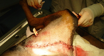
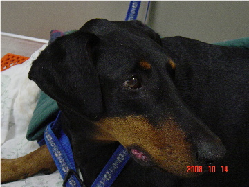
“Gibson” is a 7 year old male Doberman that presented to our clinic on the Thanksgiving weekend. His owners found him with a very large gaping wound in his groin area on the deck of their boat. They rushed him into the clinic where he was immediately stabilized by the veterinary staff on duty. He was in considerable pain and very scared. Gibson had been rescued by his current owners so this was a very stressful ordeal for him. After Gibson was stable he was placed under general anesthesia to have his wound cleaned and explored to assess the extent of the damage. He was a lucky dog since the injury didn’t extend into his abdominal cavity. He had ripped some of his muscle and done damage to his skin and fatty tissue around the laceration. Some vessels and nerves were also damaged. While under anesthesia the area was carefully put back together. With such a high area of tension and under constant motion, this was a difficult area to repair and there was concern for infection or of suture breakdown if Gibson wasn’t careful during his recovery. However, after nearly a month Gibson is doing well and has fully recovered according to his owners!
Saphire
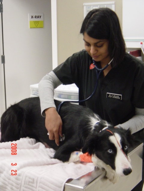
“Saphire” is a 2 year old intact female Border Collie who presented to AECFV after she vomited and then collapsed. On her initial physical exam we found that she was non-responsive, her gums where white, she had some fluid in her lungs, and there was an unusual tubular structure palpable in her abdomen. On x-rays, we found what was causing her to be so sick- she had a closed pyometra (an infection of the uterus). This condition can be fatal if it is not surgically corrected right away, however there were some concerns about taking her into surgery with the fluid in her lungs, as this could indicate a compromised heart. We performed some tests on the heart which confirmed that she did not have primary heart disease, and the decision was made to take her into surgery that night. The surgery was extremely touch and go, and she was critical for the entire night afterwards. Finally, in the morning, she started to breathe on her own and began to lift her head. Over the next few days the fluid in her lungs resolved, she appeared very bright and had a good appetite. She was sent home two days after surgery to a very happy family!
Commander
When “Commander”, a six year old two kilogram Pomeranian arrived at the clinic, he was gasping for breath as a rawhide-type chew toy got stuck in his esophagus, and was large enough to partially block his airway. His tongue was purple (never a healthy color), and he was immediately put on oxygen while the veterinarian got permission from the owner to put “Commander” under anesthesia so we could remove the toy.
It was a true emergency and a rewarding case as the doctor was quickly able to dislodge the object and “Commander” could breathe easier and much more effectively. His tongue color quickly returned to a happy, healthy pink and after recovery from his brief anesthesia, he was able to go home with no adverse consequences.
Boz
 “Boz” a 4 year old neutered male Chow Chow was presented to the Animal Emergency Clinic after having an encounter with a porcupine. Boz was in considerable distress when he arrived and had multiple quills embedded in his mouth, head, neck and forelimbs. “Boz” was given pain relief medication, anesthetized and had the quills removed. It took almost two hours to remove the quills as many of them were embedded under the skin and needed to be cut out of the tissues. The wounds were treated topically and “Boz” was discharged with antibiotics and anti-inflammatory medication.


